A virus is a unique microscopic agent that does not fit neatly into the biological package we call a cell. A cell has:
- Defined cellular structure with organelles
- Metabolic infrastructure and function – The ability to convert consumed non-living materials into biological energy and cellular components
- Homeostatic controls – The ability to regulate the internal environment
- Reproductive capacity – The ability to produce offspring
- Responsiveness – The ability to respond to external stimuli and conditions
- Capacity for growth
- Capacity for adaptation – The ability to alter form, function, or both over time in response to environmental challenges
A virus is structurally and functionally different. It is essentially an organized fusion of proteins and nucleic acids (genetic information). As such, a virus lacks:
- Defined cellular structure – Cytoplasm, a cell wall or cell membrane, ribosomes, and mitochondria are all lacking.
- Metabolic infrastructure – Without metabolic enzymes of its own, a virus is parasitic, relying on invaded cells for provision of metabolic capacity.
- Reproductive capacity – In an isolated state, a virus can’t reproduce itself and relies on the exploitation of invaded host cells to enable replication.
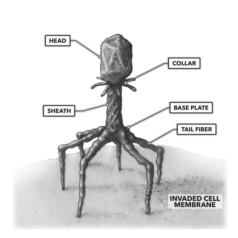
In most high school and college biology courses, we learn about the structure of a virus through the presentation of how a bacteriophage is built.

Figure 1: Bacteriophages possess a nucleic acid molecule surrounded by a protein structure, the head. The tail of the virus is composed of proteins and subdivided into the collar, sheath, base plate, and tail fibers. The tail fibers recognize complementary proteins on a target cell’s membrane and exploit those proteins to form bridging bonds with that cell.
A bacteriophage’s structure differs dramatically from other virus forms, such as the tobacco mosaic virus, the influenza virus, and the currently active SARS-CoV-2 (severe acute respiratory syndrome coronavirus 2), which drives the COVID-19 disease state (coronavirus disease originating in 2019). The bacteriophage, however, remains a good representation of the basic stages of the viral cycle:
- Adsorption – Attachment of the virus to a target cell
- Penetration – Once bound to the cell membrane, the virus transfers its nucleic acid contents into the cell to be invaded.
- Synthesis of viral proteins – The invaded cell’s internal machinery is involuntarily co-opted to produce viral proteins.
- Replication of viral nucleic acids – Instead of producing cellular genetic materials, the invaded cell replicates the viral genome.
- Packaging of viral genome and viral proteins – New viruses are assembled.
- Lysis – The invaded cell is ruptured to release newly formed viruses into extracellular spaces to amplify infection.
While these stages represent a valid generalization of how all viruses work, there are multiple types of viruses with different anatomical schemes. Each variant effects some change in the viral cycle, and each variant affects host selection and virulence. Viral categorization is most frequently done by morphology:
- Head and tail virus – These are the prototypical teaching examples; they are bacteriophages — i.e., viruses that infect bacteria.
- Filamentous virus – Many plant viruses fall into this category.
- Isometric or icosahedral virus – Poliovirus and the herpes viruses fall into this category.
- Enveloped virus – These viruses have membranes surrounding their protein shells, or capsids. Most animal viruses — including the variola virus (smallpox), human immunodeficiency virus, influenza viruses, and the coronaviruses (e.g., SARS-CoV and MERS-CoV) — fall into this category.
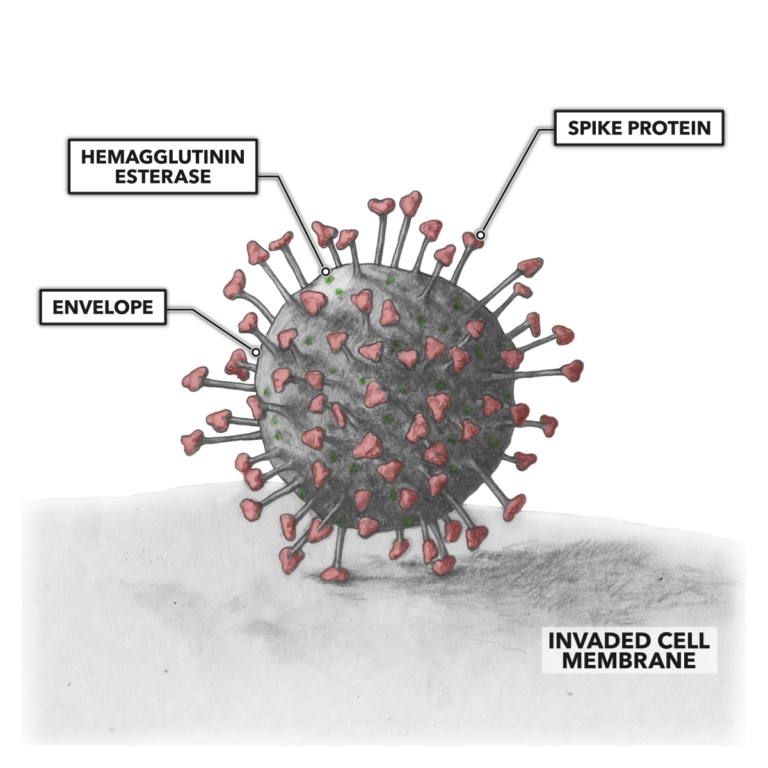
The most pressing epidemiological virus we currently face is the SARS-CoV-2 variant of the envelope virus. What makes this particular virus so insidious is not that it is dramatically more deadly than other similar viruses but that it appears to be more infectious. Biologists are currently working to understand which anatomical features of the virus create this anomalous efficiency in its entry into cells.

Figure 2: SARS-CoV-2 possesses nucleic acid materials contained within a proteinaceous envelope. Additional proteins are embedded in the envelope. Some of these proteins occur as protruding stalks with spike proteins at the terminus. These proteins recognize complementary proteins on a target cell’s membrane and exploit those proteins to form bridging bonds with that cell.
A prominent track of the rapidly evolving research into SARS-CoV-2 relates to the spike proteins of the protein capsule. These proteins interact with the cell to be invaded. Some believe targeting the spike proteins and blocking their binding capacity will provide a solution to the problem of infection. This is just one avenue scientists are investigating to get a handle on the virus and help us all return to as normal a life as possible soon.