The many ligaments of the ankle complex hold 14 bones in close proximity, including the tibia, fibula, and tarsals. The most well known of these are the lateral and medial ligaments, though some refer to them as lateral and medial collateral ligaments. The three medial collateral ligaments are better known as the deltoid ligament. The easiest and most correct method of learning and remembering the names of the ligaments is simply to use the names of the bones as prefix or suffix prompts. For example:
Tibia = tibio, tibial
Fibula = fibulo, fibular
Talus = talo, talar
Calcaneus = calcaneo, calcaneal
You can then use this convention to identify ligaments throughout the ankle and foot.

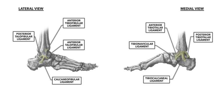
Figure 1: The lateral and medial ligaments of the ankle
Lateral Ligaments
Posterior talofibular ligament – This is the thickest and strongest of the lateral ligaments. It connects the lateral malleolus on the fibula to the posterior rim and lateral tubercle on the posterior aspect of the talus.
Anterior tibiofibular ligament – As the name indicates, this ligament attaches the lateral fibula to the anterior-medial aspect of the tibia.
Anterior talofibular ligament – This is the weakest of the lateral ligaments. It connects the anterior aspect of the lateral malleolus of the fibula to the lateral articular facet of the talus. Two-thirds of the approximately 30,000 ankle sprains reported daily in the U.S. are of the anterior talotibial ligament. When the foot is dorsiflexed and inverted at the ankle, the ligament is most vulnerable.
Calcaneofibular ligament – This ligament attaches the anterior aspect of the lateral malleolus to the lateral surface of the calcaneus about halfway along its length. In about two-thirds of the ankle sprains involving the anterior talofibular ligament, the calcaneofibular ligament is also injured.
Medial Ligaments
Tibionavicular ligament – The tibionavicular ligament attaches the medial malleolus of the tibia to the medial-posterior aspect of navicular tarsal bone.
Anterior tibiotalar ligament – This ligament connects the anterior and inferior end of the tibia to the distal and medial rim of the talus.
Posterior tibiotalar ligament – Similar to its anterior counterpart, the posterior tibiotalar ligament attaches the posterior and inferior end of the tibia to the posterior rim of the talus, just superior to the calcaneus.
Tibiocalcaneal ligament – The tibiocalcaneal attaches proximally to the point of the medial malleolus on the tibia and distally to the medial surface of the calcaneus about halfway along its length and directly below the malleolus.
The bony architecture of the medial ankle is more robust and stable than the lateral side. As such, less than five percent of all ankle ligament injuries involve medial ligaments.
Additional Reading
- Ankle Musculature, Part 1: Posterior Muscles
- Ankle Musculature, Part 2: Anterior and Lateral Muscles
To learn more about human movement and the CrossFit methodology, visit CrossFit Training.
Ankle Musculature, Part 3: Lateral and Medial Ankle Ligaments